Anatomy of the heart
The heart is a muscular, cone-shaped organ located behind the sternum and is approximately the same size as the patient’s closed fist.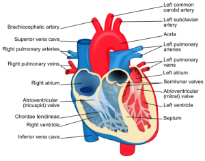
The heart walls are made up the three structures: the pericardium, myocardium and endocardium.
The pericardium is a thick fibrous membrane which surrounds the heart. Its function is to anchor the heart and prevents over distension (expanding too much). The myocardium is the central layer of the heart and is formed from cardiac muscle tissue, it is this muscle which provides the force which pumps the blood around the body.
Cardiac muscle tissue consists of a network of muscle fibres which branch and connect with each other. If one muscle fibre contracts, this contraction spreads throughout the network of muscle cells. Other structures such as blood vessels and nerves also lie within the myocardial muscle.
The endocardium provides a smooth lining inside the heart and covers the heart valves.
To allow the heart to function as a pump, valves are needed to control the flow of the blood. These are situated at the entrances and exits of the ventricles.
The valves are made up of endocardium and fibrous tissue. The tricuspid and mitral valves (which lie between the atria and ventricles) are prevented from inverting by fine tendons (chordae tendineae) attached to papillary muscles (small extensions of the myocardium).
The sinoatrial (SA) node is the heart’s natural pacemaker, containing cells that generate electrical impulses which spread through the atria, triggering contraction of the atria, forcing blood into the ventricles, and stimulating the atrioventricular (AV) node.
The AV node is linked with the atrioventricular bundle of His which transmits impulses to the left and right bundle branches and then to the Purkinje fibres which surround the ventricles. The electrical impulses travel through the Purkinje fibres causing the ventricles to contract and blood forced out of the heart.

